Friday 24 June 2011

My favourite scan so far

I popped down to the lab today to see Andy H, and was amazed by his latest scan. It's of two hand phalanges. They have fused at an angle, showing the claw hand deformity; the joint is also subluxed (partially dislocated). He kindly gave me a couple of quick screen shots, so you can see what it looks like - this will be sent off to Chris W and the texturing team next week.


I've seen so many photographs of these bones, but they've never shown the angle of fusion so well. I can't wait to see how they look when they have been cleaned up and textured!

Friday 17 June 2011

Looking after the Skeletons

One of my main jobs in Bradford is to curate our skeletal collection - which contains over 4,000 skeletons. This includes those from Chichester, some of which have been in Bradford for over 25 years! Over that time, there have been significant improvements in the way we curate skeletal remains. The first innovation in Bradford was the introduction of guidelines on 'How to Pack a Skeleton', which was already in use when I was a student here back in 1999.


One of my MSc cohort, Anwen Caffell, studied the effects of repeated handling of human remains in the collections - her work was published in 2001 in Human Remains: Conservation, Retrieval and Analysis, edited by E. Williams. She noted that the use of blue tack and masking tape damages skeletal material, and often leaves a residue behind on bones. Also, sticking broken elements back together can make them more fragile, and lead to further breakages. Perhaps the biggest concern is using material that is not labelled for teaching and research - although great care is taken that bones do not get misplaced, when it does happen it is much easier to return if there is a unique code on it! As a result of Anwen's work, and concerns of staff, many new procedures have been put in place, reducing the risk of damage to skeletal material and promoting good curatorial practice - and yes, blue tack and masking tape have been banned!

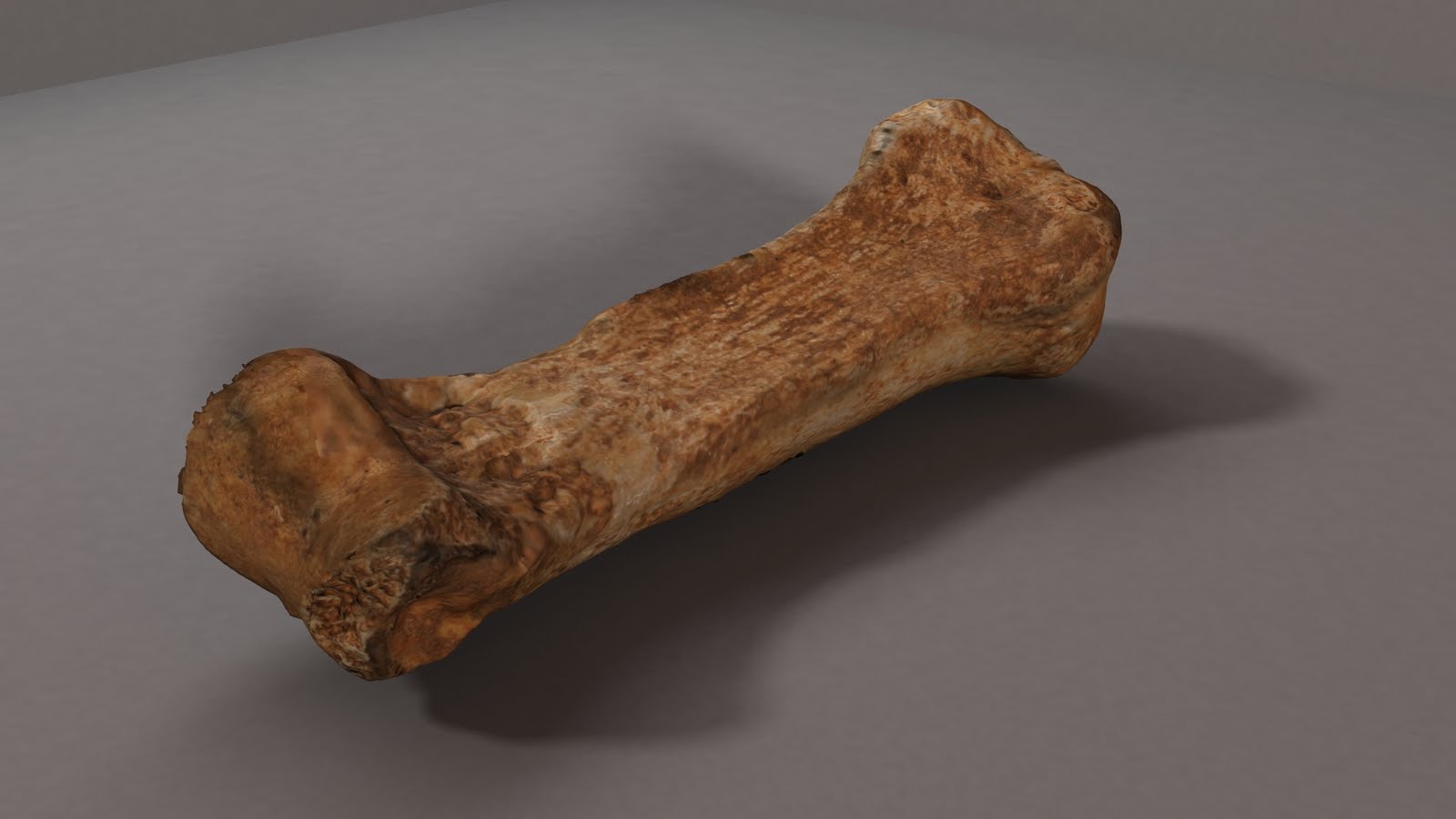
So what do these curatorial issues have to do with the digitisation project? Well the 3D scans will allow us to 'join' fragmented bones back together, or articulate bones of the hands and feet without the need of sticky substances, and handling of the bones will be reduced, as many can be examined digitally. Although there will always be a need to refer back to the original bone, the digital archive will help to preserve the collection for the researchers and students of the future.

Wednesday 1 June 2011

(Most of) our team!

As promised, here are a few pictures of the team.  Our texturing team will revealed within the next couple of weeks.


Andy Holland busy capturing point clouds.
From left-to-right: Chris Gaffney, Rebecca Swift, Tom Sparrow, Andy Wilson, Rachel Holgate, Jo Buckberry, Chris Watkins, Hassan Ugail, Andy Holland.

Group discussion


The traditional cake served up at our advisory panel meetings.

Monday 23 May 2011

Impressive results from our texturing team

In our first post, we showed you raw 3D digitised data of a phalanx bone.  What this didn't have was colour - also known as the "texture map".  For this project, we have assembled a group of 4 extremely talented animation and modelling students to work on the daunting task of texture mapping hundreds of bones. 

Tom Newbold, James Thompson, Patrick Garwood, and Toby Toochukwu have begun work on cleaning the 3D scan data, surfacing, and texture mapping.  Below are a few screen shots of a couple of the proximal phalanx bones from subject C88.


We'll be working towards standardised lighting models for the rendered videos and images, so that the colour appearance is uniform.  Below is a video of the bone shown in the last two images:


Images of the team will follow shortly.

Tuesday 17 May 2011

Scanning X-rays

Here is an update by my undergraduate placement student, Rachel, who is helping with digitising the radiographic, photographic and paper archive:


My name is Rachel Holgate and I am a placement student here at Bradford University working under Dr. Jo Buckberry, and also studying Bioarchaeology. I applied for this placement, working in BARC (Biological Anthropological Research Centre), mainly for my curiosity in bones. So far, I have thoroughly enjoyed my time here and it has cemented my interest in osteology and the hope of further study in this area.

Rachel digitising a radiograph of one of the Chichester skeletons

My involvement in the From Cemetery to Clinic project has been fantastic. Meetings, dare I say it, have been incredibly interesting and fun. So far, I have been digitising radiographs and coloured slides so that they can be accessed on the web page being created. The task I undertook was very informative from my point of view as an undergraduate student as I was able to see material first hand that is probably the best examples of clinical cases of leprosy, not to mention the opportunity to see the radiographs of the skeletal record from Chichester. I have completed digitising all the clinical radiographs and the radiographs of skeletons from Chichester that have been involved in the 3D scanning process to date. I feel incredibly lucky to be a part of this project and am thoroughly enjoying the experience so far.

Friday 6 May 2011

First Blog Updates


For our first blog entry I decided to upload our slide which we presented at the Kick-off meeting for the JISC e-content programme on 30th March.  This was a fun day, and each project representative had 3 minutes to present their project.

The slide shows one of the early bones we scanned, a proximal phalanx from the right hand of one of our subjects.  This bone is interesting because it shows the effects of  leprous claw hand which is caused by the leprosy virus attacking the nerves in the arm which results in a fixed flexion of the fingers.  The result of this is that over time a volar groove is formed - this groove can be seen near the left edge of the bone as it appears in the images:


In our next blog we'll talk about the progress of the project so far, and show some images of Andy Holland in action with the laser scanner.